Autopsies Reveal Medical Atrocities of Genetic Therapies - Dailyclout
Burkhardt Group Conclusions confirm Dr. Bhakdi's warnings
One-time or recurring donations can be made through Ko-Fi:
February 15, 2023, by Robert W. Chandler, MD, MBA
Summary:
Dr. Arne Burkhardt is one of eight international pathologists, physicians and scientists who were asked to perform a second autopsy, requested by friends and family of the deceased who were not satisfied with the results of the first autopsy.
Thirty autopsies and three biopsies were evaluated; 15 cases with routine histopathology (Step 1), three with advanced methods (Step 2), and some of the remaining 15 are included as illustrative cases.
The Step 1 group included eight women and seven men aged 28-95 (average 69).
Death occurred seven days to 180 days following the first or the second Spike-Mediated Gene Therapy (SMGT) with COMIRNATY in eight, Moderna in two, AstraZeneca in two, Janssen in one and Unknown in two.
Place of death was known in 17 cases:
Nine Non-hospital: five at home, one on the street, one in a car, one at work, one in an elder care facility
Eight Hospital: four ICU, four died having been in hospital less than two days
Special stains were used to identify Spike and Nucleocapsid Proteins, with the following differential:
COVID-19 (C-19) = + Spike + Nucleocapsid.
SMGT = + Spike – Nucleocapsid.
Causation by SMGT: Very probable in five cases, probable in seven, unclear in two and no connection in one.
Lesions were on multiple organs including: Brain, Heart, Kidney, Liver, Lungs, Lymph Node, Salivary Gland, Skin, Spleen, Testis, Thyroid and Vascular.
Lymphocyte Infiltration, present in 14 of 20 cases (70%), was a common feature and involved multiple organs. Case 19 had at least five different organs involved. CD3+ Lymphocytes were dominant.
The Vascular System was targeted by Lymphocyte Infiltration in seven (35%) of the cases and included sloughing endothelium, destruction of the vessel wall, hemorrhage and thrombosis.
A condition called Lymphocyte Amok was described by Dr. Burkhardt: Lymphocyte accumulation in non-lymphatic organs and tissues that might develop into lymphoma.
Five cases of unknown foreign material in blood vessels were identified. The favored explanation for origin of this material was aggregated Lipid Nanoparticles (LNPs).
Multiple pathologic processes were involved: Apoptosis, Coagulopathy, Clotting/Infarction, Infiltration/Mass Formation, Inflammation, Lysis, Necrosis and Neoplasia.
Röltgen, et al. https://www.cell.com/cell/fulltext/S0092-8674(22)00076-9found that COVID-19 depleted Lymphatic Germinal Centers (LGCs) whereas SMGT stimulated them, suggesting a possible origin of “Hunter/Killer” CD3+ Lymphocytes that are attracted to certain tissues, particularly the vascular system.
An expanded program of autopsy following SMGT is recommended in order to further understand the actions of SMGTs and to help formulate new treatments for the constellation of pathology associated with such drugs.
Burkhardt Group Conclusions:
Histopathologic analyses show clear evidence of vaccine-induced autoimmune-like pathology in multiple organs.
That myriad adverse events deriving from such auto-attack processes must be expected to very frequently occur in all individuals, particularly following booster injections.
Beyond any doubt, injection of gene-based COVID-19 vaccines place lives under threat of illness and death.
We note that both mRNA and vector-based vaccines are represented among these cases, as are all four major manufacturers.
Histopathology
This report is the first in a series in which harms from the Lipid Nanoparticle (LNP) Messenger Ribonucleic Acid (mRNA) therapeutics and other Spike-mediated products will be examined from the point of view of the pathologist, a medical doctor that studies specimens obtained from removal of tissue from living persons, bulk resection or biopsy, or after death. Such examinations make or confirm a diagnosis and provide a basis to determine causation of tissue mass or cause of death. Histopathology refers to the study of abnormal tissues.
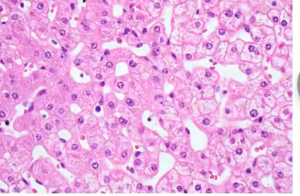
Tissues are examined using careful inspection of specimens with the naked eye followed by examination by light microscopy employing a variety of different stains to highlight important features of cells, tissues and organs. A common stain used is hematoxylin and eosin, H & E for short, which stains nuclei blue, cytoplasm pink or red, collagen fibers pink and muscles red.
Many of the photomicrographs in this and subsequent articles will have been stained with H & E. Pathologists display sections prepared with H & E along with the magnification used, such as 40 times (40X) or 100 times (100X) magnification.
Read the full report on dailyclout.io
Burkhardt Group Conclusions Confirm Dr.Bhakdi's Warnings
Related articles:
Dr. Michael Yeadon: THIS MUST STOP! Pfizer Documents Show FDA Knew of Death Risk
Dr. Michael Yeadon: The Most Important Single Message I’ve Ever Written
PREMEDITATED MASS MURDER: Alarming Data From Canada and Vaccines Batch Scandal


Crime Against Humanity doesn’t even begin to cover this.
NIH also does lots of genetic experiments to help Big Pharma produce more drugs. There are very few Big Pharma drugs that don't require supplement drugs to counter or alleviate negative symptoms from the first drug.